Friday, 19 June 2020

Review of Terahertz Pulsed Imaging for Pharmaceutical Film Coating Analysis

Alves-Lima, Décio, Jun Song, Xiaoran Li, Alessia Portieri, Yaochun Shen, J. Axel Zeitler, and Hungyen Lin. "Review of Terahertz Pulsed Imaging for Pharmaceutical Film Coating Analysis." Sensors 20, no. 5 (2020): 1441.

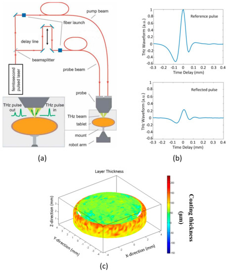
Terahertz pulsed imaging (TPI) was introduced approximately fifteen years ago and has attracted a lot of interest in the pharmaceutical industry as a fast, non-destructive modality for quantifying film coatings on pharmaceutical dosage forms. In this topical review, we look back at the use of TPI for analysing pharmaceutical film coatings, highlighting the main contributions made and outlining the key challenges ahead.

Thursday, 18 June 2020

Nondestructive measurement of mill-scale thickness on steel by terahertz time-of-flight tomography

Zhai, Min, Alexandre Locquet, Cyrielle Roquelet, Patrice Alexandre, Laurence Daheron, and D. S. Citrin. "Nondestructive measurement of mill-scale thickness on steel by terahertz time-of-flight tomography." Surface and Coatings Technology (2020): 125765.

AbstractFig. 1. Optical photographs of the three scale films of thickness (a) 28

We measure in a nondestructive and noncontact fashion the thicknesses of three scale films with thicknesses 28.5 ± 1.4 μm, 13.4± 0.9 μm, and 5.1 ± 0.3 μm on steel substrates employing terahertz time-of-flight tomography combined with advanced signal-processing techniques. Wüstite is the dominant phase in the scale films, though magnetite and hematite are also present. Because wüstite is electrically insulating, the incident terahertz electromagnetic pulses largely penetrate into the scale film; however, the pulses are entirely reflected by the underlying electrically conductive steel substrate. Because the film layers are thin, in some cases optically thin, the distinct pulses reflected at the air/scale and scale/steel interfaces overlap in time and thus are not visually evident in the reflected terahertz signal, necessitating the use of deconvolution techniques to recover the sample structure. We compare the merits of three deconvolution techniques, one unsuccessful (frequency-wavelet domain deconvolution) and two successful (sparse deconvolution and autoregressive extrapolation), to characterize the thicknesses of these scale films.

ExperimentalThe THz experiments were carried out in an air -conditioned laboratory at 22 o C with humidity < 48 %. Otherwise, the samples were stored in a dry box to retard the transformation of wüstite from transforming into magnetite. The experiments shown here were conducted within the 3 months of sample fabrication to ensure that the scale films were primarily composed of wüstite. The THz reflection measurements are taken on a TeraView TPS Spectra 3000, a pulsed, broadband THz time -domain spectroscopy (TDS) system, shown in Fig. 4. 

Wednesday, 17 June 2020

Terahertz Imaging and Characterization Protocol for Freshly Excised Breast Cancer Tumor

Vohra, Nagma, Tyler Bowman, Keith Bailey, and Magda El-Shenawee. "Terahertz Imaging and Characterization Protocol for Freshly Excised Breast Cancer Tumors." Journal of Visualized Experiments 158 (2020).

This manuscript presents a protocol to handle, characterize, and image freshly excised human breast tumors using pulsed terahertz imaging and spectroscopy techniques. The protocol involves terahertz transmission mode at normal incidence and terahertz reflection mode at an oblique angle of 30°. The collected experimental data represent time domain pulses of the electric field. The terahertz electric field signal transmitted through a fixed point on the excised tissue is processed, through an analytical model, to extract the refractive index and absorption coefficient of the tissue. Utilizing a stepper motor scanner, the terahertz emitted pulse is reflected from each pixel on the tumor providing a planar image of different tissue regions. The image can be presented in time or frequency domain. Furthermore, the extracted data of the refractive index and absorption coefficient at each pixel are utilized to provide a tomographic terahertz image of the tumor. The protocol demonstrates clear differentiation between cancerous and healthy tissues. On the other hand, not adhering to the protocol can result in noisy or inaccurate images due to the presence of air bubbles and fluid remains on the tumor surface. The protocol provides a method for surgical margins assessment of breast tumors.

Application of Terahertz Spectroscopy and Imaging in the Diagnosis of Prostate Cancer

Zhang, Ping, Shuncong Zhong, Junxi Zhang, Jian Ding, Zhenxiang Liu, Yi Huang, Ning Zhou, Walter Nsengiyumva, and Tianfu Zhang. "Application of Terahertz Spectroscopy and Imaging in the Diagnosis of Prostate Cancer." Current Optics and Photonics 4, no. 1 (2020): 31-43.

Abstract

The feasibility of the application of terahertz electromagnetic waves in the diagnosis of prostate cancer was examined. Four samples of incomplete cancerous prostatic paraffin-embedded tissues were examined using terahertz spectral imaging (TPI) system and the results obtained by comparing the absorption coefficient and refractive index of prostate tumor, normal prostate tissue and smooth muscle from one of the paraffin tissue masses examined were reported. Three hundred and sixty cases of absorption coefficients from one of the paraffin tissues examined were used as raw data to classify these three tissues using the Principal Component Analysis (PCA) and Least Squares Support Vector Machine (LS-SVM). An excellent classification with an accuracy of 92.22% in the prediction set was achieved. Using the distribution information of THz reflection signal intensity from sample surface and absorption coefficient of the sample, an attempt was made to use the TPI system to identify the boundaries of the different tissues involved (prostate tumors, normal and smooth muscles). The location of three identified regions in the terahertz images (frequency domain slice absorption coefficient imaging, 1.2 THz) were compared with those obtained from the histopathologic examination. The tissue tumor region had a distinctively visible color and could well be distinguished from other tissue regions in terahertz images. Results indicate that a THz spectroscopy imaging system can be efficiently used in conjunction with the proposed advanced computer-based mathematical analysis method to identify tumor regions in the paraffin tissue mass of prostate cancer.


Terahertz Imaging and Spectroscopy Data Acquisition 
All prostate tissue were tested using a TPI system (TeraPulse 4000, TeraView, Cambridge, England) operating in Reflection Spectrum and Imaging Module (RIM). This module adopts a single-point scanning imaging method. A raster-scanning system that changes the position of the sample through a mobile station in an X-Y plane was used, which allowed the terahertz radiation to pass through the individual points of the sample to be measured. The maximum size of the sample that the system can scan is 20 mm * 20 mm, and the minimum scanning step is 0.1 mm. The axial resolution of our TPI system used is around 30 microns and the maximum lateral resolution is around 250 microns. In this study, the scanning area of the sample was about 15 * 5 mm, the resolution of each point was 0.2 mm, and the diameter of the terahertz spot at the focal point is 0.2 mm

Monday, 15 June 2020

Laser process of transparent conducting surfaces for terahertz bandpass ultrathin metamaterials.

Wang, Qinghua, Bingtao Gao, Fatima Toor, Mark Arnold, and Hongtao Ding. "Laser process of transparent conducting surfaces for terahertz bandpass ultrathin metamaterials." In Laser-based Micro-and Nanoprocessing XIV, vol. 11268, p. 112680H. International Society for Optics and Photonics, 2020.

Abstract
Terahertz (THz) optoelectronics have great potentials in communication, imaging, sensing and security applications. However, the state-of-the-art fabrication processes for THz devices are costly and time-consuming. In this work, we present a novel laser-based metamaterial fabrication (LMF) process for high-throughput fabrication of transparent conducting surfaces on dielectric substrates such as quartz and transparent polymers to achieve tunable THz bandpass filtering characteristics. The LMF process comprises two steps: (1) applying ultrathin-film metal deposition, with a typical thickness of 10 nm, on the dielectric substrate; (2) creating periodic surface pattern with a feature size of ~100 microns on the metal film using nanosecond pulsed laser ablation. Our results demonstrate the LMF-fabricated ultra-thin metal film exhibits newly integrated functionalities: (a) highly conductive with sheet resistance of ~20 Ω/sq; (b) optically transparent with visible transmittance of ~70%; (c) tunable bandpass filtering effect in the THz frequency range; and (d) extraordinary mechanical durability during repeated fatigue bending cycles. The scientific findings from this work will render an economical and scalable manufacturing technique capable of treating large surface area for multi-functional THz metamaterials.


for full paper see https://www.spiedigitallibrary.org/conference-proceedings-of-spie/11268/112680H/Laser-process-of-transparent-conducting-surfaces-for-terahertz-bandpass-ultrathin/10.1117/12.2544054.short

Experimental

… 2.5 THz Transmission Measurement Experimental THz transmission spectra, were generated using THz-Time Domain Spectroscopy (THz-TDS) technique 52,53. A 1000D TeraView spectrometer (TeraView Limited, Cambridge)